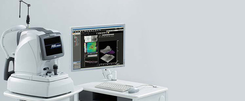
Ocular Coherence Tomography (OCT) is arguably one of the most clinically useful diagnostic instruments ever invented. It allows detailed cross-sectional images of structures of the eye in the same way an MRI scan does in other parts of the body. It is used to image all parts of the eye including the cornea, iris, lens, vitreous, retina and choroid.
Lakkis Optometry have recently introduced OCT Angiography (OCT-A) which can now assess active blood flow in the arteries and capillaries of the retina. Unlike traditional angiography, it does not require an injection of fluorescent dye into the body. It is particularly useful in wet macular degeneration and diabetic retinopathy.
The results of the OCT scan are available for immediate review and interpretation. Your optometrist will discuss the results with you and advise of any treatment or referral that may be required.


